Percutaneous Atrial Septal Defect Closure (ASD & PFO)

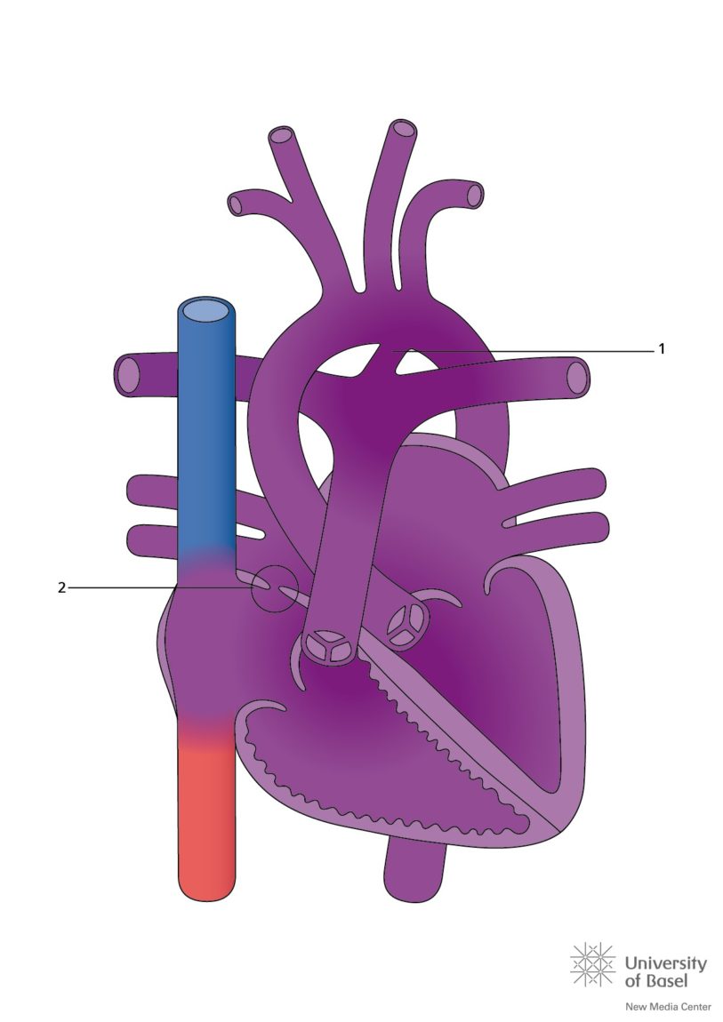
The septum is the wall separating the right side of the heart from the left side. The atrial septum divides the right and left atria and the ventricular septum divides the right and left ventricle. In a fetal heart, the foramen ovale, a small hole located in the atrial septum, allows blood to bypass the fetal lungs and travel directly to the rest of the body. This is because the fetus gets oxygen from the umbilical cord, not the fetal lungs. The foramen ovale usually closes within days after birth due to pressure changes in the infant’s chest. If the foramen ovale does not close, it is called a patent foramen ovale (PFO).

An atrial septal defect is an abnormal opening in the septal wall between the two upper chambers of the heart called the atria. This opening can cause mixing of arterial (oxygen rich) blood with venous (oxygen poor) blood and may create extra work for the right side of the heart. PFO is like a flap whereas ASD is a hole in the atrial septum that may allow blood to flow to the lungs in larger quantities.

Symptoms from an ASD may include: shortness of breath with exertion, fatigue, rhythm problems of the heart such as atrial fibrillation, rarely patients may have symptoms of a stroke.  Most patients have no symptoms but your doctor may find an enlarged heart or high pressure in the lungs due to increased lung blood flow.

If the ASD is of significant size, it may cause overload to the right heart and lungs causing enlargement as well as elevation of right heart and high lung pressures which can reduce lifespan and increase the risk for rhythm problems of the heart and can cause disability and reduction in functional capacity.  IF an ASD is causing right heart volume overload it should be closed either percutaneously or surgically.

A catheter based (non-surgical) option involves the insertion of a device called the Amplatzer septal occlude (ASO) device across any ASD to close it like a clamshell to stop flow across your ASD and thereby reduce the flow from your left heart upper chamber to your right heart upper chamber.  Various companies make devices to close an ASD. We most commonly use the Abbott Septal occlude device or the Gore ASD Cardioform device. This device is constructed with a braided nitinol (nickel + titanium) mesh containing a cloth like mesh to encourage clot formation and closure of the ASD.

Repairs

Sometimes, depending on the type of ASD and PFO, the hole can be closed without open heart surgery. This is called a percutaneous repair and it is done in the cardiac catheterization lab. The procedure will typically take 1-2 hours to complete and may require the use of general anesthesia, meaning you will be asleep and won’t remember the procedure.

ABBOTT Septal Occluder
GORE ASD Cardioform Occluder

The surgical approach involves open heart surgery to stich or patch close the ASD communication between the two upper chambers of the heart.

Patent Foramen Ovale Closure

Symptoms related to a PFO may include stroke (or mini-stroke) with no other clear identifying cause, shortness of breath (due to mixing of non-oxygenated blood from the right side of the heart with oxygenated blood in the left side of the heart), decompression illness (from deep diving), and in some cases, migraines with aura.

If your symptoms are deemed related to a PFO, you may undergo a transesophageal echocardiogram (ultrasound with tube in the esophagus to see the heart closely and a right and left cardiac catheterization (to check blood oxygen levels +/- arteries of the heart).  Once accepted, the procedure is usually performed with general anesthesia, but in some cases, under conscious sedation. A catheter is placed through your vein and a clamshell device placed to close the PFO (similar to an ASD).  The devices we use are the Abbott Talisman Occluder or the Gore Cardioform Septal Occluder, both made out of a nitinol frame.

Abbott Talisman
GORE Cardioform Septal Occluder

What to expect?

Pre-procedure: you will be educated about the procedure in the pre-procedural clinic by one of our nurses.  You will be instructed on arrival times, when to stop eating the night before and which medications to take.  Bloodwork will be performed prior to the procedure.  Your doctor will arrange for you to have a special test called a transesophageal echocardiogram, a special ultrasound involving a same day admission to image your ASD better and to rule out other heart defects or blood clot in the upper chambers of the heart.  Your ultrasound doctor will explain this procedure in detail to you on the day of the procedure.

Day of the procedure:  Your doctors will explain the procedure to you again and obtain informed consent. The procedure will then be performed in the Y-2 Cardiac Cath Labs under conscious sedation or general anesthesia, depending on your particular situation you may need general anesthesia and a trans-esophageal echocardiogram to monitor your procedure.  Alternatively, this may be done using a device called an intra-cardiac echocardiogram without the need for general anesthesia.  Both groins will be cleaned and sometimes an arm artery may be prepared and cleaned.  Usually two veins and one artery will be accessed with small tubes called sheaths to allow monitoring with the intracardiac echo, to monitor blood pressures and lastly to perform the delivery of the ASO device.  The procedure may last from 1.5 – 2 hours.  The tubes from the legs will then be removed either in the lab or in the holding area.  Bedrest will be for generally at least 4-6 hours post procedure.  The physicians will already have initiated aspirin 81-325 mg once a day and likely clopidogrel 75 mg once a day (for a period of six months) prior to or the day of your procedure.  It is to be expected that you will remain in hospital overnight to obtain an ultrasound (echocardiogram) of your heart the next morning and for further observation. If you have had a PFO closure, you may go home either the same evening or the next day based upon some specific criteria as adjourned by the treating physicians. However, your physician may decide that you need to stay more or less than this depending on procedure complexity and complications.

Post Procedure: You will be informed by your physician for clinic follow-up or will be called at home shortly after discharge.  You will be given appointments for routine echocardiogram (heart ultrasound) monitoring.  Usually these will be at 3 months and 1 year, and then yearly thereafter.  You will be instructed and be given prescriptions for aspirin and clopidogrel antiplatelet medications to be taken daily without missing doses at all.  You will be given instructions regarding antibiotic prophylaxis for a period of 6 months for all dental and other relevant procedures you may have over the 6 months post procedure.  It is also advised to avoid heavy activity, strenuous activity, and heavy lifting for one month.

This device is MRI-compatible at 1.5 Tesla and is conditional at 3.0 Tesla.  You should not have an MRI for the 6 weeks following implantation

SYMPTOMS TO WATCH FOR POST PROCEDURE, POST DISCHARGE:

  1. Chest pain – call 911 to come to hospital
  2. Sudden weakness, loss of speech, sudden change in vision, numbness – call 911 to come to hospital
  3. Dizziness, palpitations, fainting or lightheaded spell – call 911 to come to hospital
  4. Fever, bleeding at leg site, pain or swelling at leg site worse than before discharge – call 911 to come to hospital

RISKS / COMPLICATIONS OF CATHETER BASED ASD CLOSURE:

  1. Erosion of device into other heart chambers or into the lining of the heart – 1/500, may manifest as chest pain, sudden cardiac death, dizziness – call 911, this complication most commonly happens in the first 72 hours, but has been reported to happen out to 3-5 years, this will usually require open heart surgery to correct
  2. Rhythm problems – <1-2%, sometimes requiring medications, blood thinners and less commonly but possibly permanent pacemaker insertion
  3. Device embolization or movement of device to unintended location during or after procedure – approximately 1% – may be retrievable with catheters or may need open heart surgery to retrieve the device and close the ASD subsequently
  4. Vascular (blood vessel) complication or rupture, sometimes requiring surgery or blood transfusion – 1-2%
  5. Stroke or mini-stroke or clot formation on the device – <1%
  6. Headaches/migraines – uncommon but may occur or persist post device, generally treated conservatively
  7. Fever / device infection – <1%
  8. Allergic reaction to the dye/contrast – <1%
  9. Hemolysis – breakdown of blood due to blood crossing device, generally self limiting, <1%
  10. Cardiac perforation – may occur during procedure or later due to device erosion as above, <1%, often needs open heart surgery to correct
  11. Heart Valve damage – uncommon <1%
  12. Transient chest pain or small heart attack, mini stroke due to air escaping to the heart arteries or brain. <1%
  13. Death – <1%
  14. Bleeding or allergic reaction to aspirin or clopidogrel used to prevent clot outside the device